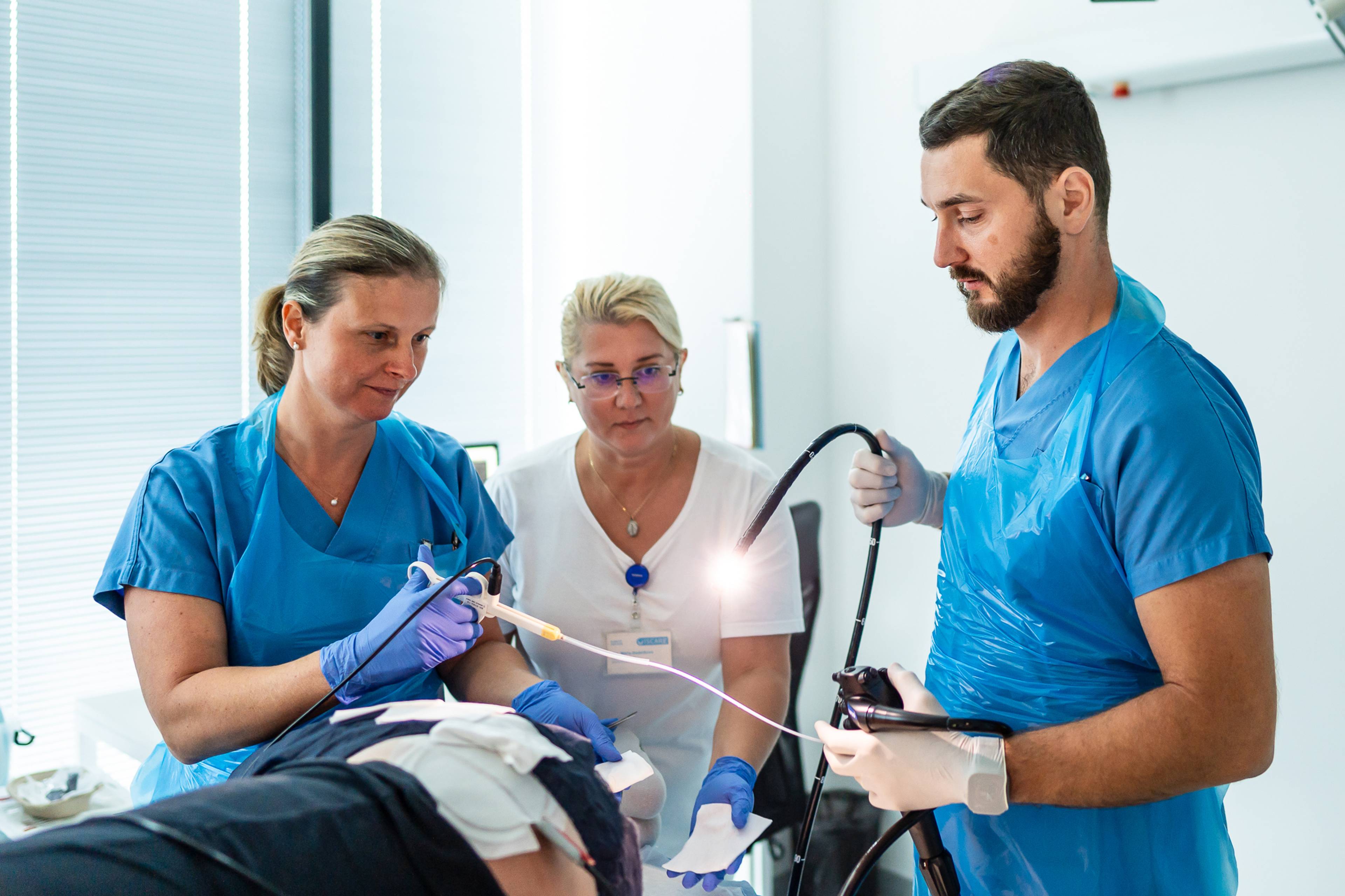
Capsule Enteroscopy: Painless imaging of the intestinal tract
- Capsule enteroscopy is a painless imaging examination of the small intestine performed using a special diagnostic capsule. This capsule, slightly larger than a standard vitamin capsule (11x26 mm), is equipped with a microcamera, light source, and transmitter.
- The patient swallows the capsule, which takes color images of the inside of the intestine as it passes through the digestive tract. The images are continuously recorded by a recorder that the patient carries with them. After passing through the intestine, the capsule is excreted naturally with the stool within a few days (maximum two weeks).
- Before the examination itself, it is necessary to undergo an ultrasound, CT/MRI of the intestines, and a colonoscopy. These preliminary examinations serve to detect any narrowing of the intestine and minimize the risk of subsequent complications with the passage of the capsule. Only after obtaining these results can the doctor request approval for reimbursement from the health insurance company. We do not perform examinations solely at the client's request – a doctor's referral is always required.
- This type of examination does not allow for the collection of tissue samples for further examination and does not allow for any therapeutic interventions. Capsule enteroscopy is performed on an outpatient basis.
Preparation for the examination:
1 day before the examination:
- Follow a residue-free diet.
- The evening before the examination, take ½ dose of a bowel cleansing agent recommended by your doctor or pharmacy specialists, e.g. ISCARE.
On the day of the examination:
- Do not eat after midnight, but drinking is allowed.
- Take your morning medication 2 hours before the examination.
Take a look
What it looks like
Gastroenterology ISCARE


