Ultrasonography of the abdomen and intestines
Sonography is an examination of the abdomen and intestines with a special probe to assess the activity and extent of inflammatory changes in patients with IBD.
Ultrasonography of the abdomen and intestines
Abdominal Sonography
Sonography is an examination of the abdomen using a special probe, whereby the parenchymal organs of the abdominal cavity – the liver, bile ducts, gallbladder, pancreas, kidneys, spleen, vascular structures, and organs of the small pelvis, i.e. the bladder, female reproductive organs, and prostate in men – are examined through the abdominal wall. In some patients, the examination includes liver elastography, which measures the degree of stiffness (fibrosis) of the liver tissue caused by the accumulation of connective tissue in the liver.
Intestinal Sonography
A special probe is used to examine the intestinal loops through the abdominal wall in order to assess the activity and extent of inflammatory changes and possible complications in patients with IBD. The examination is also used when acute inflammation of the diverticula or appendix is suspected.
Preparation for the examination:
- The day before the examination, it is advisable not to eat foods that cause flatulence – excessive amounts of gas in the digestive tract impair the patient's examinability.
- The patient should be fasting – do not eat for 6 hours before the examination, but may drink a small amount (approx. 200 ml) of still water.
- The patient should not urinate for 2 hours before the examination of the small pelvis and intestines.
Examination procedure:
A small amount of transparent gel is applied to the skin of the abdomen, and then the organs are examined by moving the probe over the abdominal wall. The examination begins with the patient lying on their back, and during the examination, the patient may be asked to change position to their left or right side. Sometimes, for a better evaluation of the findings, it is necessary for the patient to hold their breath for a moment while inhaling. The examination is painless, has no contraindications, and is safe even for pregnant patients.
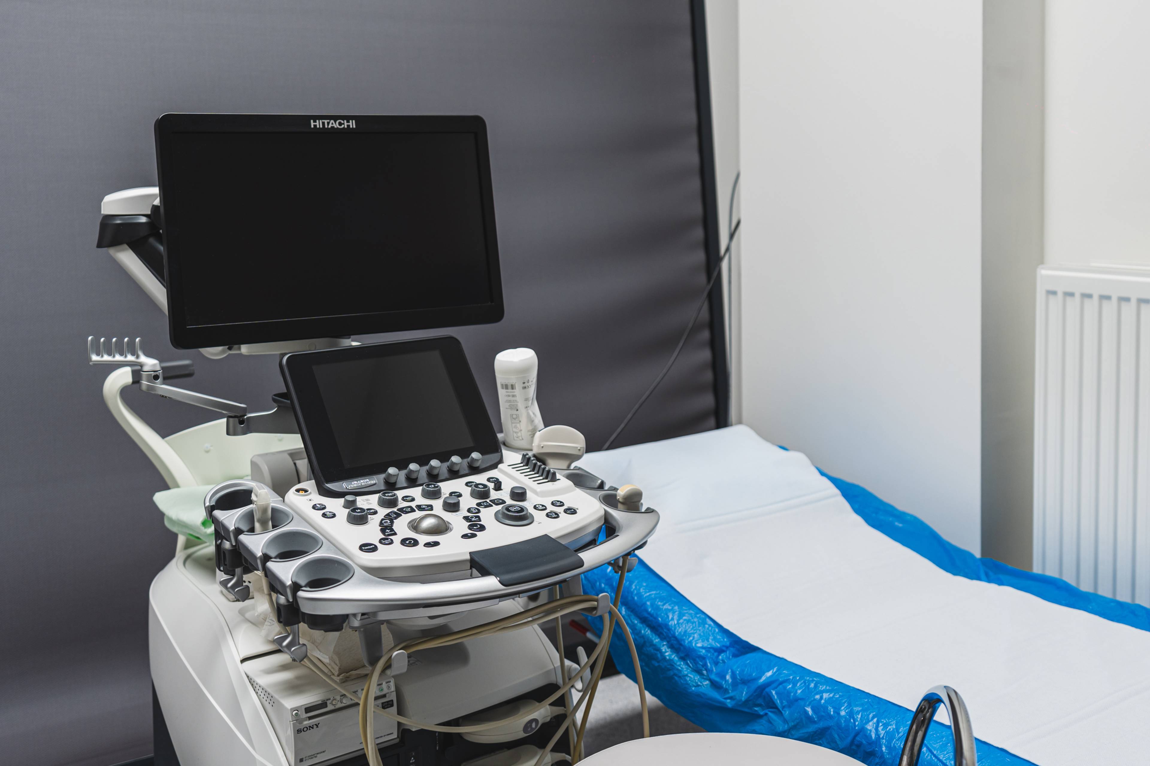
Take a look
What it looks like
Gastroenterology ISCARE